VERTEBRAL COLUMN
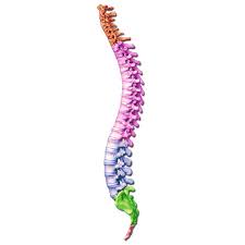
Vertebral column is made by 26 bones.
24 separate vertebrae extend downwards from the occipital
bone of the skull;then there is the sacrum,formed from five fused vertebrae,and
lastly the coccyx,or tail,which is formed from between three to five small
fused vertebrae, and lastly the coccyx, or tail, which is formed from between
three to five small fused vertebrae.

The vertebral column is divided into
different regions.
The first seven vertebrae, in the neck, form the cervical
spine; the next twelve vertebrae are the thoracic spine and the next five the
lumbar spine, the lowest vertebra of which articulates with the sacrum.
Each
vertebra is identified by the first letter of its region in the spine, followed
by a number indicating its position. For example, the topmost vertebra is
called C1, and the third lumbar vertebra is called L3.
The movable vertebrae have many characteristics in common,
but some groups have distinguishing features.
CHARACTERISTICS OF A TYPICAL VERTEBRA

THE BODY.
The body
of each vertebra is situated anteriorly.
The size varies with the site.
They
are smallest in the cervical region and become larger towards the lumbar
region.
THE VERTEBRAL
(NEURAL) ARCH.
This enclosed a large vertebral foramen.
It is the area
behind the body, and forms the posterior and lateral walls of the vertebral
foramen.
The lateral walls are formed from plates of bone called pedicles, and
the posterior walls are formed from laminae.
Projecting from the regions where
the pedicle meets the lamina is a lateral prominence called transverse process,
and where the two laminae meet at the back is a process called the spinous
process.
These are the bony prominences that can be felt through the skin along
the length of the spine.
The neural arch has four articular surfaces; two
articulate with the vertebra above the two with the one below.
The vertebral
foramina form the vertebral (neural) canal that contains the spinal cord.
REGION-SPECIFIC VERTEBRAL CHARACTERISTICS
CERVICAL VERTEBRAE

These are the smallest vertebrae.

The transverse processes
have a foramen through which a vertebral artery passes upwards to the brain.
The
first two cervical vertebrae, the atlas and the axis, are typical.
The first cervical vertebra, the atlas, is the bone on which
the skull rests.

Below the atlas is the axis, the second cervical vertebra
(C2).
The atlas is essentially a ring of bone, with no distinct
body or spinous process, although it has two short transverse processes.
It possesses
two flattened facets that articulate with the occipigtal bone; these are
condyloid joints and they permit nodding of the head.

The axis sits below the atlas, and has a small body with a
small superior projection called the odontoid process (also called the dens, meaning
tooth).
This occupied part of the posterior foramen of the atlas above, and is
held securely within it by the transverse ligament.
The head pivots (i.e. turns
from side to side) on this joint.
The seventh cervical vertebra C7, is also known as the
vertebra prominens.
It possesses a long spinous prominence terminating in a
swollen tubercle, which is easily felt at the base of the neck.
THORACIC VERTEBRAE.

The thoracic vertebrae are larger than the cervical
vertebrae because this section of the vertebral column has to support more body
weight.

The bodies and transverse process have facets for articulation with the
ribs.
LUMBAR VERTEBRAE.

These are the largest of the vertebrae because they have to
support the weight of the upper body. They have to support the weight of the
upper body.
They have substantial spinous processes for muscle attachment.
SACRUM.

This consists of five rudimentary vertebrae fused to form a
triangular or wedge-shaped bone with a concave anterior surface.
The upper
part, or base, articulates with the 5th lumbar vertebra.
On each
side it articulates with the ilium to form a sacroiliac joint, and at its
inferior tip it articulates with the coccyx.
The anterior edge of the base, the
promontory, protrudes into the pelvic cavity.
The vertebral foramina are
present, and on each side of the bone there is a series or foramina for the
passage of nerves.
COCCYX

This consists of the four terminal vertebrae fused to form a
very small triangular bone, the broad base of which articulates with the tip of
the sacrum.
FEATURES OF THE VERTEBRAL COLUMN
INTERVERTEBRAL DISCS

The bodies of adjacent vertebrae are separated by
intervertebral discs, consisting of an outer rim of fibrocartilage (annulus
fibrosus) and a central core of soft gelatinous material (nucleus pulposus). They
are thinnest in the cervical region and become progressively thicker towards
the lumbar region, as spinal loading increases.
The posterior longitudinal
ligament in the vertebral canal helps to keep them in place.
They have a
shock-absorbing function and the cartilaginous joints they form contribute to
the flexibility of the vertebral column as a whole.
INTERVERTEBRAL FORAMINA.

When two adjacent vertebrae are viewed from the side, a
foramen formed by a gap between the vertebral pedicles can be seen.
Half of the
wall is formed by the vertebra above, and half by the one below.
Throughout the length of the column there is an
intervetebral foramen on each side between every pair of vertebrae, through
which the spine nerves, blood vessels and lymph vessels pass.
LIGAMENTS OF THE
VERTEBRAL COLUMN.

These ligaments hold the vertebrae together and keep the
intervertebral discs in position.
The transverse ligament maintains the odontoid process of
the axis in the correct position in relation to the atlas.
The anterior longitudinal ligament extends the whole length
of the column and lies in front of the vertebral bodies.
The posterior longitudinal ligament lies inside the
vertebral canal and extends the whole length of the vertebral column in close
contact with the posterior surface of the bodies of the bones.
The ligament flava connect the laminae of adjacent
vertebrae.
The ligamentum nuchae and the supraspinous ligament connect
the spinous processes, extending from the occiput to the sacrum.
CURVES OF THE VERTEBRAL COLUMN.

When viewed from the side, the vertebral column presents
four curves: two primary and two secondary.
The fetus in the uterus lies curled up so that the head and
the knees are more or less touching.
This position shows primary curvature.
The
secondary cervical curve develops when the child can hold up his head (after
about 3 months) and the secondary lumbar curve develops when he stands upright
(after 12 to 15 months).
The thoracic and sacral primary curves are retained.
MOVEMENTS OF THE
VERTEBRAL COLUMN
The movements between the individual bones of the vertebral column
and very limited. However, the movements of the column as a whole are quite
extensive and include flexion (bending forward), extension (bending backward),
lateral flexion (bending to the side) and rotation. There is more movement in
the cervical and lumbar regions than elsewhere.
FUNCTIONS OF THE VERTEBRAL COLUMN
These include the following:
·
Collectively the vertebral foramina form the
vertebral canal, which provides a strong bony protection for the delicate
spinal cord lying within it.
·
The pedicles of adjacent vertebrae form intervertebral
foramina, one on each side, providing access to the spinal cord for spinal
nerves, blood vessels and lymph vessels.
·
The numerous individual bones enable a certain
amount of movement.
·
It supports the skull.
·
The intervertebral discs act as shock absorbers,
protecting the brain.
·
It forms the axis of the trunk, giving
attachment to the ribs, shoulder girdle and upper limbs, and the pelvic girdle
and lower limbs.
share us on :munikrishnadn.blogspot.com


0 Comments